Sheffield scientists uncover never before seen pictures of deadly bacteria in fight against superbugs
and live on Freeview channel 276
The new findings about antibiotic resistant bacteria could help scientists fight against the rise in superbugs.
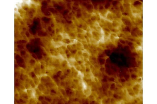
Scientists captured never before seen, high-resolution images of the cell wall structure of the deadly bacterium Staphylococcus aureus – more commonly known as MRSA.
Advertisement
Hide AdAdvertisement
Hide AdThe images showed a new and unexpected structure of the outer bacterial layers of the resistant bacterium MRSA – which overturned previous theories about how bacteria grow and antibiotics work.

The research, which was funded by UK Research and Innovation (UKRI), the Wellcome Trust and the White Rose University Consortium, gives new insight into the composition of the bacterial cell wall and will inform future studies about how antibiotics can be developed to combat antibiotic resistance.
Previous studies of the cell wall in any organism do not have a comparable resolution or molecular scale.
PhD Researcher from the University of Sheffield’s Department of Physics and Astronomy, Laia Pasquina Lemonche said: “Many antibiotics work by inhibiting the bacteria’s production of a cell wall, a strong but permeable skin around the bacteria which is critical for its survival.
Advertisement
Hide AdAdvertisement
Hide Ad“We still don’t understand how antibiotics like penicillin kill bacteria, but this isn’t surprising because until now we had remarkably little information about the actual organisation of the bacterial cell wall.
“This study provides that essential stepping stone which we hope will lead to both a better understanding of how antibiotics work and to the future development of new approaches to combat antimicrobial resistance.”
The team used an advanced microscopy technique to produce the images – where a sharp needle is used to feel the shape of the surface and build an image at the scale of individual molecules. Professor Jamie Hobbs, Professor of Physics at the University of Sheffield, said: “It is by physicists and biologists working together that we've been able to make these breakthroughs in our understanding of the bacterial cell wall.”Researchers are now using the same techniques to understand how antibiotics change the architecture of the cell wall and how such changes influence antimicrobial resistance.